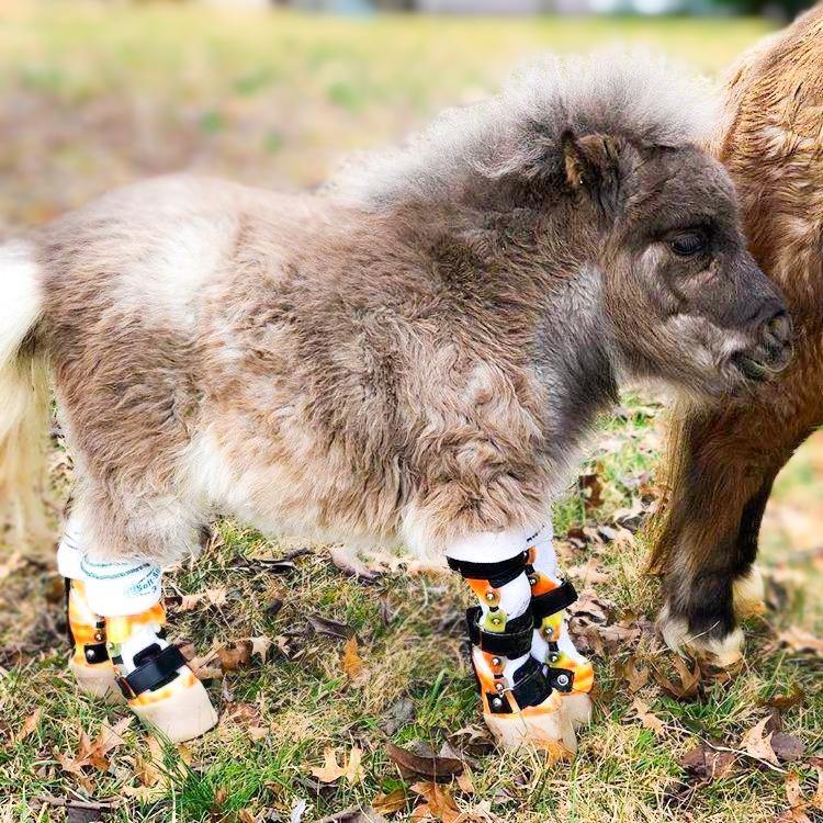
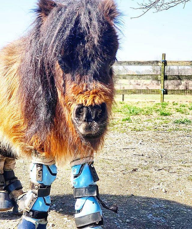
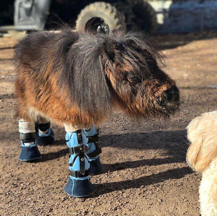
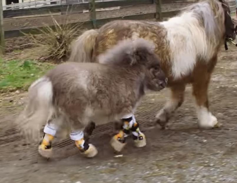
Custom Equine Leg Brace | Horse Leg Brace
$900.00


Equine Custom Brace Order Process

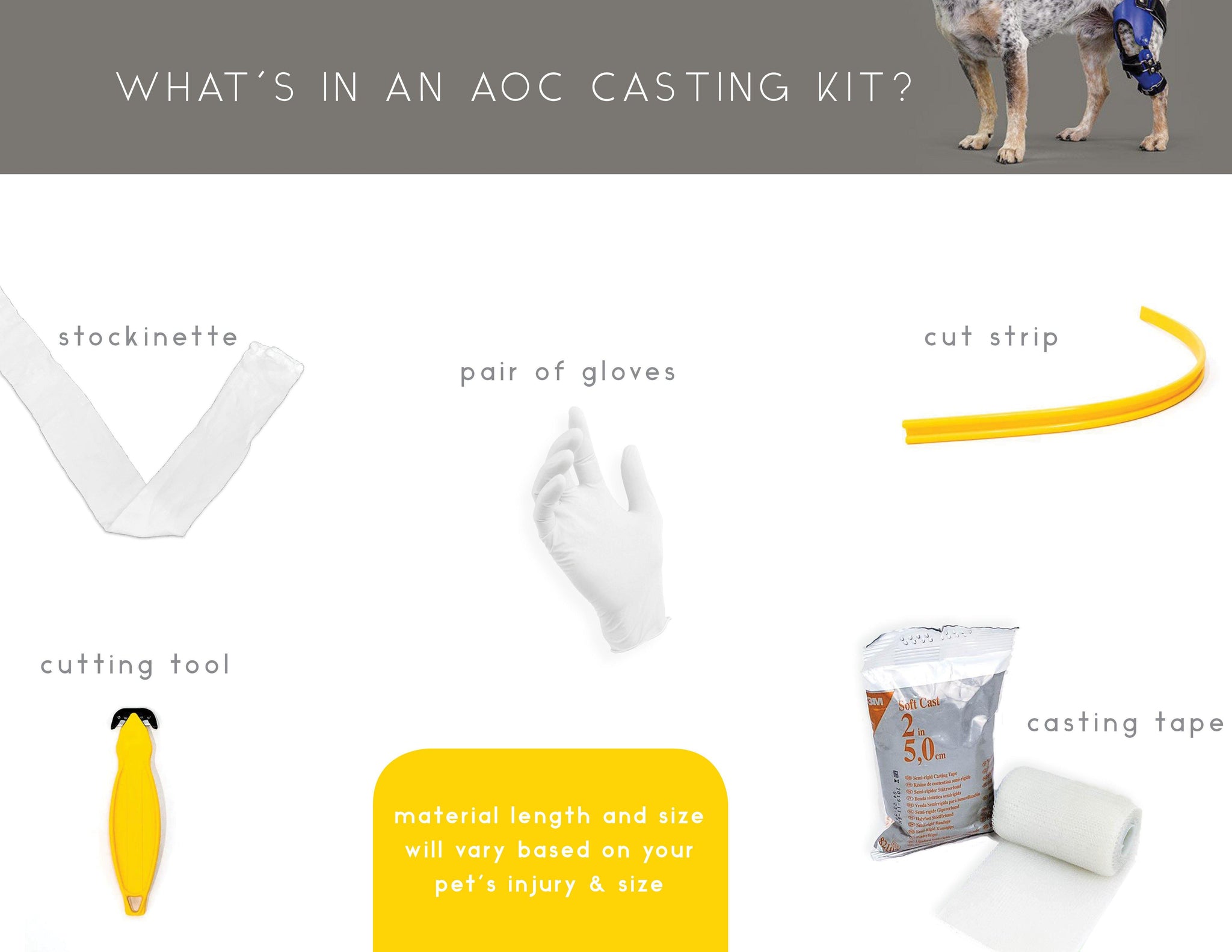
Cast
We send you a Casting Kit for you to make a mold of your horse's limb.

FIT
We ship you the finished custom brace. You fit it to your horse. Your horse is on their way to recovery!
BUILDING YOUR Horse's custom BRACE
AFTER RECEIVING YOUR Horses'S CAST,
FABRICATION TIME WILL TAKE 2-4 weeks.
Benefits
- Option for non-surgical candidates due to age, cost, other contraindications
- Stabilizes the joint
- Provides support to the affected area
- Decreases healing time and decreases cost
- Return to improved activity level and near normal joint function
- Allows for easier wound inspection
- Releases pressure on Contra-lateral Limb
conditions
- A relatively common fracture in racehorses, thought to occur from recurrent "micro-damage" during training that finally gives way
- Presents as a longitudinal crack down the lower end of the cannon bone, splitting the cannon bone into the fetlock joint surface
- In flat racing horses, the crack usually occurs on the left forelimb, on the outside (lateral) side of the bone
- The fracture also occurs in Standardbred racehorses and in those horses also commonly occurs on a hind limb
- Articular fractures cause an added problem, damaging the joint surface, resulting in arthritis
- The injury usually results in very severe lameness but occasionally causes less severe lameness
- There is usually swelling of the lower limb and fluid filling of the fetlock joint
- Stabilizes the joint
- Provides support to the affected area
- Decreases healing time and decreases cost
- Return to improved activity level and near normal joint function
- Allows for easier wound inspection
- Release pressure on Contra-lateral Limb
- Forearm fractures can occur as a single (radius or ulna only) or combined (both bones) fracture
-
When both bones are fractured at different levels and there is a joint injury at the wrist or elbow, these are described as Galeazzi or Monteggia fractures
- Galeazzi facture: Most often a displaced fracture in the radius and a dislocation of the ulna at the wrist, where the radius and ulna come together
- Monteggia fracture: Most often a fracture in the ulna and the top (head) of the radius is dislocated at the elbow joint
- Fractures of the tibia, which is the weight-bearing bone of the leg, are often associated with fracture of the fibula
- Displaced fractures usually involve both the tibia and fibula
- The skin and subcutaneous tissue over the anterior and medial tibia are very thin and therefore lower leg fractures are often open
- Even in closed fractures, the soft tissue can become compromised. The fibula is well covered by soft tissue except at the lateral malleolus
- Tibial fractures in adults are usually caused by direct blows or falls on to the tibial shaft
- Spiral fractures of the tibia and fibula may be caused by violent twisting injuries, usually from contact sports
- The fibula is fractured in 75-85 % of cases with fractures of the tibia
- Flexor Tendon Laxity usually occurs in newborn foals, but can occur in slightly older foals
- This laxity can range from a slight drop in the fetlock to the fetlock(s) actually touch the ground
- Flexor tendon laxity is common in premature or dysmature foals
- Mild laxity usually resolves on its own as the foal gets stronger and exercises, often within a few days to one week
- If the laxity is more pronounced, then hoof trimming to create a flat, weight-bearing surface is typically undertaken
- Flexural contractures often are referred to as “contracted tendons,” occurring when the affected foal stands, it appears that the tendons are tense and too short. Also, the deformity can be present at birth (congenital) or develop in the older foal (acquired)
- The source of this problem in the newborn is not completely understood, but is thought to be caused by malposition of the foal within the uterus
- Additionally, nutritional abnormalities and even genetics have been implicated as some mares produce multiple foals with flexural deformities
- Flexural contracture results in the flexion of the joints of the lower limb(s)
- Joints most commonly affected are the carpus, fetlock, and coffin joint, with one or multiple joints or legs affected
- Flexural deformities also can occur in older foals, known as an acquired flexural deformity
- Mild cases of flexural deformities can resolve on their own with light bandages and exercise, while Moderate cases might need splinting, bracing and/or casting with the hoof exposed, called tube casting
- These types of contractures typically occur in older foals and in fairly specific locations
- Young foals (one to six months) might develop contracture at the coffin joint, while Older foals (at least three months as well as yearlings and occasionally 2-year-olds) might develop contracture at the fetlock joint
- These contractures occur in the forelegs, usually in both (bilateral), except for cases where the contracture in one leg is due to lack of use because of pain
- Acquired flexural deformity of the coffin joint often is referred to as “club foot”
- The foot can vary from a dished appearance with the heel raised to a boxy shape with the hoof wall nearly perpendicular to the ground
- In very severe cases, the foal or horse might walk on the front (dorsal) aspect of the hoof or fetlock
- Mild cases might require only a decrease in nutrition; in young foals weaning might work
- Angular limb deformities are deviations that occur from side to side, such as when the leg deviates from the carpus, tarsus, or fetlock to the outside (laterally) or inside (medially)
- A lateral deviation is called a valgus deformity and a medial deviation is called a varus deformity
- These deviations are extremely common and can be congenital (present at birth) or acquired (develop later in the foal’s life)
- The Collateral Ligament of the hock consists of long and short groups medially and laterally that run at almost ninety degrees to each other, providing significant stability in a wide range of positions
- Affected horses usually present with hindlimb lameness of variable severity that gets worse with exercise
- Poor performance
- Chronic or acute rear limb lameness
- Lameness worsens with exercise
- Swelling, thickening and pain of the soft tissues surrounding the affected area
- Shortened stride length
- Toe dragging
- The carpus is composed of three joints: the antebrachiocarpal (radiocarpal) joint, the middle carpal (intercarpal) joint, and the carpometacarpal joint
- Injury to the carpus most commonly occurs within the antebrachiocarpal and middle carpal joints
- High-energy injuries, such as falls or kicks, to the carpus cause acute single load failure. The resultant injury often causes open or comminuted fractures and carpal instability
- Single load injuries including luxations of one or more carpal joints are less common than those developing as a result of repetitive loading
- Injury to the intercarpal ligaments can occur and usually involves the palmar medial intercarpal ligament within the middle carpal joint
- Injury to the collateral ligaments is usually associated with avulsion fractures of the proximal aspect of the vestigial metacarpal bones or distal radius
- Injury to the extensor tendons usually develops in association with severe flexural deformities or lacerations
- Injuries to the palmar aspect of the carpus do occur, but they are less common than dorsal injuries and have been associated with hyperextension or hyperflexion of the carpus during recovery from anesthesia
- The Suspensory Ligament supports the fetlock and protects it from hyperextension (i.e., dropping too low) at exercise
- The ligament begins at its attachment to the back of the upper cannon bone in both the fore and hindlimbs. It runs downwards close to the back of the cannon bone before dividing into two branches each of which attaches to one of the sesamoid bones at the back of the fetlock. Some fibers continue and attach to the upper pastern area.
- The ligament and its branches are strong but only slightly elastic
- Excessive stress can occur to the ligament when a horse lands after a jump or when it travels at fast speeds
- Essentially, an over-stretching injury can occur resulting in damage to the ligament
- The damage may be only slight tearing of fibers at the level of their 'origin' (upper cannon bone) or their 'insertion' (sesamoid bones)
- Damage is usually accumulative over a period of time and may be considered a type of 'repetitive strain injury'
Features
- Custom-fitted to your horse's limb
- Non-porous medical grade foam cushions and eases pressure on the affected area
- Durable hook and loop strap system allows easy adjustments for fit and comfort
- Made in the USA
- Removable
- Two-month warranty for brace alterations and modifications
- One-year warranty for manufacturing defects
- Lifetime warranty on Tamarack Hinges
- Free replacement straps during the first 90 days
why an aoc custom brace?


technology
- Hand-crafted using vacuum forming and thermal technology
- Waterproof
- Tamarack hinges allow motion that mimics normal ligament mobility
- Tamarack-Tether hinges allow natural ligament motion while preventing hyper-extension and hyper-flexion
- A specialized ROM hinge can be fabricated with the brace instead. There is an additional $200 charge when fabricating a custom brace that requires a ROM hinge
how to cast
The type of cast that will be required for us to make your horse's custom brace will depend on your horse's diagnosis. Our fabricators will determine what type of cast is needed and we will send you specific instructions with your casting kit.
MY CASTING DIDN'T GO WELL
CARE:
- Use light soapy water or antibacterial wipes to clean the plastic shell and non-porous liner of the brace.
- Hand-wash pads as needed and let them air dry.
- Remove any hair or dirt from the hook and loop straps with a wire brush, gently brushing in one direction on the hook side only.
USE
- It is highly recommended that you follow the break-in period protocol for your horse. This helps to prevent irritation and allows your horse to become comfortable when wearing their brace. Failure to follow the break-in period could result in further injury to your horse.
- Your horse may wear their brace whenever they are active. Custom Equine braces are waterproof and therefore can be used in the water, such as for hydrotherapy sessions.
- We recommend that you remove your horse's brace when they are not active for extended periods of time. This allows their leg to relax, prevents irritation, and allows their skin breathe.
- Ideally, the brace should not be worn for more than 14 hours a day as this could cause irritation. However, if your veterinarian recommends that your horse wear the brace for more than 14 hours a day then you may do so but only with frequent, thorough leg and brace examinations. Please check your horse's leg and brace at least 2-3 times in a 24 hour period after the initial break-in period is complete.
BREAK-IN PERIOD
Similarly to breaking in a new pair of shoes, the Custom Equine Brace should initially be worn in short increments as your horse builds up a tolerance to the device. For best results, place the brace on your pet for only half an hour the first day. You can typically increase this time by a further half hour each subsequent day. You can facilitate this process by positively reinforcing and rewarding your horse for wearing the brace. Please keep in mind that the break-in period and time of each increment will depend on your pet's diagnosis and overall acceptance of the brace.
Please note that this break-in period is highly recommended but not required if your veterinarian recommends otherwise for the health of your horse. If you do not follow our recommended break-in period, we require that you check your pet's skin and brace frequently and thoroughly, at least 6 times per day in a 24 hour period for the first two weeks. Once your horse has successfully completed the break-in period without irritation occurring, we recommend that you check your horse's skin and brace 2-3 times a day in a 24 hour period.
If there is any irritation or skin breakdown, discontinue use of the brace for one full day. If irritation continues, contact us at care@aocpet.com.
SKIN & BRACE EXAMINATION
It is crucial that you thoroughly inspect your pet's skin after each use of the Custom Equine Brace. This is especially important during the initial break-in period. While it is not uncommon to see minor problems for the first few weeks of brace usage, make note of any blisters or other unusual marks on the skin. Skin redness that does not disappear within an hour after the brace is removed and other signs of skin breakdown are indications of excessive pressure from the brace. The brace can easily be modified to relieve these areas of irritation.
You should also carefully examine the brace itself after each use. Identify any cracks, tears, or areas of unusual wear. Padding and the hook and loop straps can be replaced by contacting Animal Ortho Care. Consult our warranty terms for other details on the service process.
HYGIENE & CLEANING
Both the Custom Equine Brace and areas of skin that come into contact with it must be kept clean. This reduces the chance of your horse contracting contact dermatitis or other skin conditions. Regularly clean the brace with a mixture of half water, half isopropyl alcohol. After cleaning, dry with a towel or let the brace dry at room temperature. Do not speed the drying process through use of applied heat, such as from a hair dryer. Never put a wet brace onto your pet, as it will affect the fit. A properly-fitted brace can be worn in water, but should be thoroughly dried after use before re-application.
MATERIALS:
- Outer Shell: polypropylene, polyethylene, co-polymer
- Inner Liner: closed-cell polyethylene foam
FAQS
WHAT IS INCLUDED WITH THE PURCHASE OF A CUSTOM equine BRACE?
Purchases of our Custom Equine Braces include a free casting kit, which you will use before we begin fabricating the brace itself. Once you have cast your horse's leg - or legs if you are ordering a pair of Custom Braces - you will mail us the casts. At that point we craft your brace order based on the cast of your horse's leg, and then send the Custom Braces back to you.
WHAT HAPPENS IF THE BRACE DOES NOT FIT CORRECTLY?
Please contact us and we will work with you to find the fit! The hook and loop straps give you some freedom to adjust the brace yourself. Depending on the issues with the fit, we may instruct you to ship it back to us for more extensive modification.
Please note it is imperative that you take an accurate cast of your horse's leg in order for the brace to have the best possible fit. Please refer to our casting directions and be sure that you are familiar with them before attempting a casting.
WHAT IF MY horse's BRACE NEEDS REFURBISHMENT?
Refurbishing your horse's brace is easy. Strap replacements are free within the first 90 days of your purchase if you order and replace them yourself. We offer various methods for refurbishing your brace depending on what needs to be done. You can order new Straps (1 inch or 1.5 inch) and Pads to replace on your own. If you don't want to replace the straps yourself or need a liner replacement, you can use our Custom Brace Refurbish Service and we will refurbish the brace based on what you order.
HOW OFTEN CAN MY horse WEAR THIS BRACE?
There is a two-week break-in period where you will either gradually increase the time your horse wears their Custom Equine Brace by 30 minute increments OR you will check your horse's brace at least 6 times in a 24 hour period. Once this period is complete, you may leave the brace on your horse for up to 14 hours a day.
Please note that this brace can be worn all day if your veterinarian recommends this for the health of your pet. You will need to check your horse's skin and brace 2-3 times per day to ensure there is no irritation. We recommend that you remove the brace when your horse is resting, this will prevent irritation and allow their leg to air out.
Check for any signs of irritation and/or swelling after removing the brace.
HOW LONG IS THE FABRICATION PROCESS?
Once we receive the cast of your horse's leg, it takes 2-4 weeks to fabricate the brace.
HOW LONG WILL SHIPPING TAKE?
We offer free 5-7 business day ground shipping in the US for both the casting kit and custom brace, as well as 2 Business Day or Overnight shipping options for an additional charge. Once we receive the cast of your horse's leg and fabricate the Custom Brace, we will ship that using the same option you selected at checkout.
IS THE CASTING PROCESS DIFFICULT?
The difficulty of casting will depend on your horse; more anxious or badly injured horse's may find the process more distressing. It is recommended that you perform the process with at least 3-4 people depending on your horse's energy level.
Detailed instructions on how to cast are included in the casting kit. We can walk you or your veterinarian through the casting process, so please contact us with questions or concerns before you begin casting your horse's leg. We also offer FREE on-site casting at our Arden Hills, Minnesota location.
Net Orders Checkout
| Item | Price | Qty | Total | |
|---|---|---|---|---|
| Subtotal |
$0.00 |
|||
| Shipping | ||||
| Total | ||||